3 Axis Tomofraphey Analitical Holder

  • HATA TEM holder top

3-axis orientation alignment Tomography
High efficiency EDS Analysis
Easy to link with other tools by using cartridges

Accurate 3D visualization (By courtesy of:Prof. Hata, Prof. Mitsuhara )

HPT-Cu

tomography tem holder

Austenitic steel

tomography tem holder

AL-Mg-Si

tomography tem holder
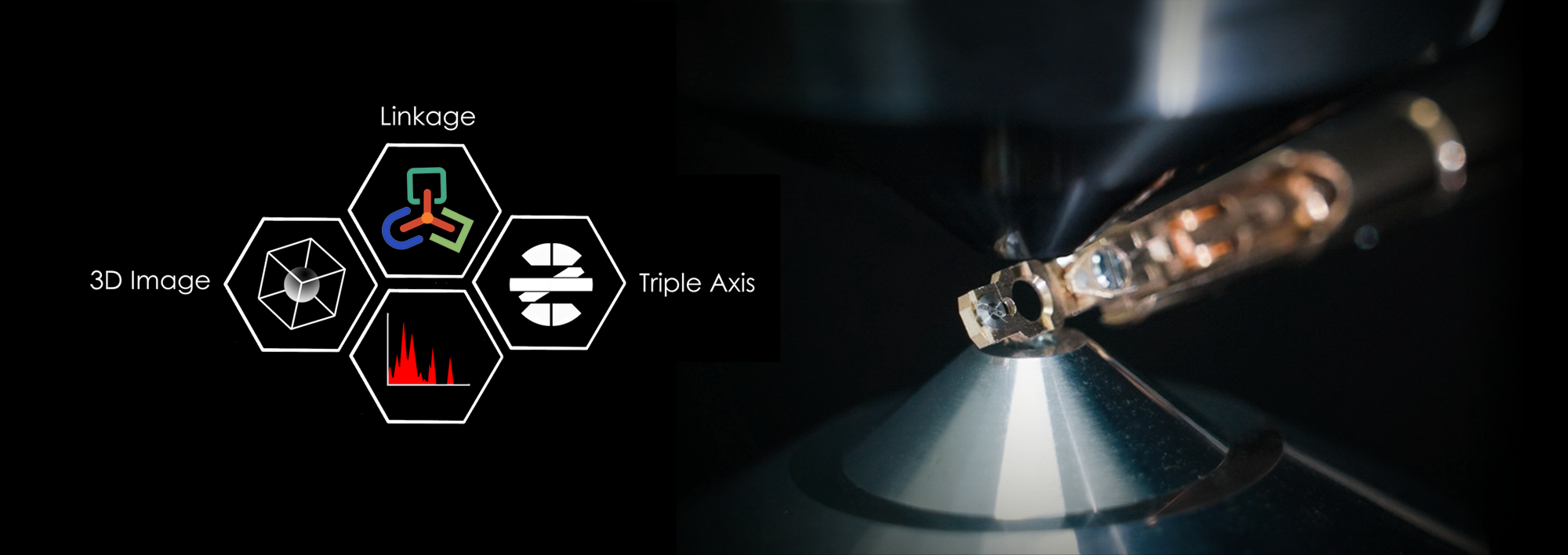
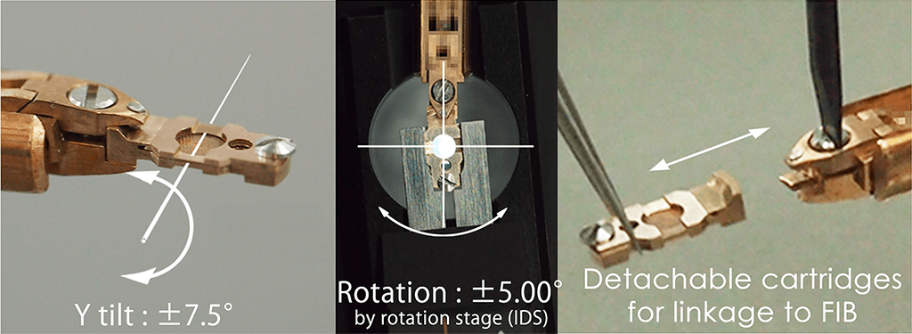
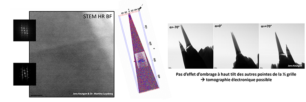
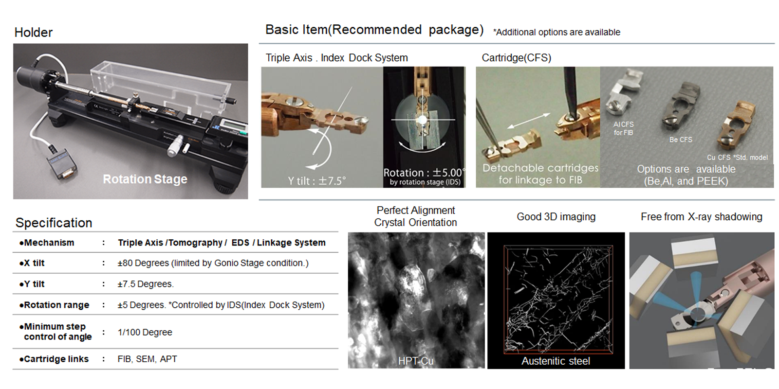
In general, the crystal orientation is aligned by two tilt axis(X tilt + Y tilt or rotation),but X axis tilting, the crystal orientation is getting off axis.
Therefore, it is necessary for perfect alignment to apply not only Y tilting but also specimen rotation, it can be tilted at a high angle on perfect aligned crystal orientation, and a beautiful 3D construction is possible by this tem holder system.

- Multi Function Structure -

  • tomography tem holder

- High detection efficiency structure for EDS analysis -

Free form X-ray shadowing

  • tomography tem holder

EDS detection efficiency is improved by the geometry of TEM Holder

  • tomography tem holder

High Resolution EDS analytical performance test:Column mapping of SrTiO3

  • tomography tem holder

- Double tilt Cartridge Linkage System -

The concept of our TEM holder is to make it into a cartridge.

  • tomography tem holder

- Comparison Tomography between TEM and Atom probe Tomography -

  • tomography tem holder

- Needle Probe Cartridge-
By creating a cartridge of our TEM Holder compatible with the Needle probe,
we have made it possible to perform tomography even with narrow gaps such as UHR.

  • HATA Holder

TEM Holder Specification

  • HATA Holder Spec

High-Angle Triple-Axis tem holder

Applicable instrument:
TFS and JEOL TEM
Max X (α) tilt range +/-80° ( Limited by the Gonio stage tilt angle. )
Max Y (β) tilt range +/- 7.50° Supports high-resolution
Double tilt + cartridge structure

Sample rotation Stage (IDS)

            Maximum Rotation range +/-5.00°
            ( Controlled by Mel-Build IDS )
           Note: Not in-situ rotation
           Minimum step control of angle:0.01°
           Illumination for specimen